Tissue Clearing & Expansion Microscopy Methods for Medical Application
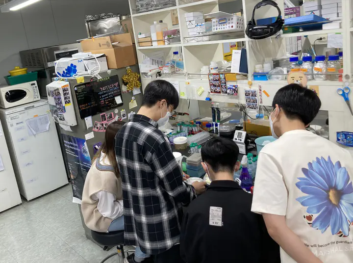 Image credit: Seung Hyun Ryu
Abstract
3D Tissue clearing is the technique that enhanced 3D volume imaging resolution with many approaches: delipidation, decolourization, decalcification, and refractive index(RI) matching.
Expansion microscopy(ExM), a nanoscale imaging technique with a conventional fluorescent microscope makes it easier to get super-resolution images. It uses water-swellable polymer expanding samples(as 4x - 20x in each dimension) with deionized water.
Today, I will show you how to subculture Cos-7 cells, how to stain nuclear with SytoX(Green fluorescent nucleic acid stain), how to transfer protein signals to polymer gels, and how to expand polymer gels as 10x and image ExM. We also analyze samples verifying expansion ratio with many proven methods.
(Kitae Kim shows how to clear and immunostain tissues with various methods and how to 3D imaging with the light-sheet microscope.)
Course Outline: This subject can provide broad perspectives on medicine for students by covering multidisciplinary areas. In addition, the students can experience the application of basic medical knowledge and realize the importance of basic medical sciences. The students can select the specific subject according to their interest and can learn the subject consistent with their knowledge level.