Expansion Microscopy(ExM) Imaging Method
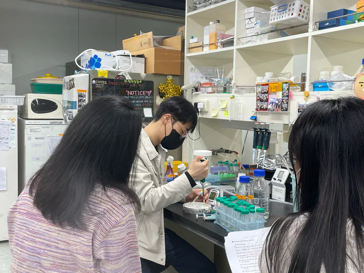 Image credit: Seung Hyun Ryu
Abstract
How small scale do you want to image? There are many super-resolution imaging techniques like STORM, STED, PALM… However, those techniques require expert skills and equipment to get one high-resolution image.
Expansion microscopy(ExM), a nanoscale imaging technique with a conventional fluorescent microscope makes it easier to get super-resolution images. It uses water-swellable polymer expanding samples(as 4x - 20x in each dimension) with deionized water.
Today, Kitae Kim and I will show you how to immunostain β-tubulin on Cos-7 cells, transfer protein signals to polymer gels, expand polymer gels, and image ExM. We also analyze samples verifying expansion ratio with many proven methods.
Date
Mar 26, 2022
Location
Seoul National University College of Medicine
103 Daehak-ro, Jongno-gu, Seoul 03080
Click on the Slides button above to view the built-in slides feature.